Call Us : +44 (0)1424 444633
Call Us : +44 (0)1424 444633
In 1965, Goldman documented the earliest report of tattoo pigment interaction with short-pulsed lasers.[18] He compared the reaction of a dark blue tattoo to a QSRL with nanosecond pulses to that of a microsecond-pulsed ruby laser. He found nonspecific thermal necrosis with microsecond impacts, while nanosecond impacts only produced transient edema accompanied by a peculiar whitening of the impact area, which lasted about 30 minutes. No thermal necrosis was present, but tattoo fragments remained in the dermis. The mechanism of this reaction was unknown but not believed to be thermal because of normal measurements taken with a thermistor. Because retention of tattoo pigment was reported, this modality was originally interpreted as a failure. However, Goldman followed the patient's progress and noted continued fading of the treated area.
In 1992, Goldman communicated that he was unable to continue this work because his engineer had been fatally electrocuted, and the project was abandoned. Three years later, other investigators confirmed and expanded these results by using a QSRL (694 nm, 20 ns) to remove blue and black tattoo pigment successfully without tissue damage.[19] Biopsies performed after 3 months showed absence of tattoo pigment and no evidence of thermal damage. These effects were dose dependent, with fluences of 5.6 J/cm2 or less showing absence of thermal damage but incomplete pigment removal. Higher fluences led to subepidermal blistering similar to a second-degree burn and dermal fibrosis at 3 months, although pigment removal was more complete. Subsequent studies concluded that this treatment modality was impractical because of the small target areas and the risk of coagulation necrosis of tissue surrounding the tattoo pigment.
Reid et al continued to study the QSRL, and, in 1983, they published an additional report on the removal of black pigment in professional and amateur tattoos. They reported good results, particularly with amateur tattoos but noted several disadvantages, including the need for multiple treatments (often 6 or more) for complete pigment removal. They also reported scarring in 2 patients and emphasized the need to use relatively low powers (but above the threshold to produce immediate tissue whitening) and the importance of the treatment interval. At least 3 weeks between treatments were necessary for tissue healing and pigment removal by macrophages. In vitro tests showed an immediate reaction in the ink that could be explained only by a chemical reaction.
Biopsy specimens revealed evidence of ink fragmentation into smaller particles, which were then phagocytized by macrophages. These 2 phenomena correspond with clinical observations of an immediate reduction in visible pigment in the first week after treatment, followed by gradual fading over the next several weeks without further therapy.
Dose Response
Stimulated by studies that found that the QSRL preferentially damaged melanized cells in animal skin and by the tattoo studies previously cited, researchers at the Wellman Laboratories of Photomedicine at Harvard Medical School completed a detailed dose-response study of the QSRL in 1990
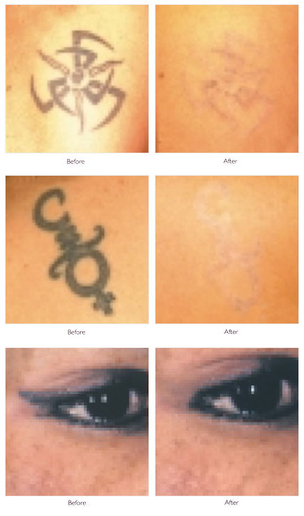
By using a 40- to 80-nanosecond pulse, they treated 41 tattoos 5 times at doses of 1.5-4 J/cm2. Of 27 amateur tattoos, 8 cleared at a rate greater than 75% (all treated at 4 J/cm2) in an average of 2.6 treatments. None of the 14 professional tattoos reached this level within 5 treatment sessions, and frequent purpura and rare, punctate bleeding occurred. Twenty-eight tattoos with residual pigment were treated up to 5 times at fluences of 5-8 J/cm2 with more tissue damage, frequent purpura, and occasional superficial erosions noted. At the completion of this phase, more than 75% clearance was achieved in an additional 10 amateur tattoos (average of 3.5 treatments) and 3 professional tattoos (single treatment).
The overall results were excellent in 78% of amateur tattoos but in only 23% of professional tattoos. Despite these discouraging statistics, the authors were optimistic that the QSRL would become the preferred treatment for tattoos because of the rare result of scarring. In addition, the authors stressed that the competitive absorption by melanosomes, which led to vacuolization of melanocytes and keratinocytes, with hypopigmentation noted in 39% (low fluences) to 46% (higher fluences) of patients. Normal pigmentation was reestablished progressively over a 4- to 12-month period; however, 4 of the 10 tattoos examined 1 year after treatment still showed confettilike hypopigmentation.
The authors agreed with previous investigators that the immediate whitening represents rapid, localized heating with steam or gas formation, resulting in dermal and epidermal vacuolization. An unexplained brief emission of white light as the laser pulse struck the tattoo pigment was noted. Interestingly, histologic persistence of altered tattoo pigment in areas of clinical clearing was noted, suggesting a change in the optical properties of the tattoo pigment.
This study did not establish an optimal treatment interval. Although the mean interval was 3 weeks, intervals of 1-5 weeks were used with no statistical difference in response. Blue and black pigments were most responsive, whereas green and yellow responded less well and red was poorly responsive to the red wavelength (694 nm). Purpura and punctate bleeding most likely represent indirect vascular injury from photoacoustic waves generated by the laser's interaction with tattoo pigment.
Results
Scheibner et al reported preliminary results of treatment of 101 amateur and 62 professional tattoos using fluences of 2-4 J/cm2 with a 40-nanosecond pulse width, spot sizes of 5 and 8 mm, and a treatment interval of 5-6 weeks. Although response details are lacking, after an average of 4 treatments, 87% of amateur tattoos were more than 80% clear, while of 62 professional tattoos, only 11% were more than 80% clear. Treated areas required 10-14 days to heal, remained erythematous for an additional 1-3 weeks, and developed hypopigmentation lasting 2-6 months in most patients. Skin textural changes resolved over 6-8 weeks, with no scarring reported. Tattoos of the face and the neck responded faster but were more sensitive to tissue damage, necessitating lower fluences. A better response was noted with older, professional tattoos and blue inks in comparison to other colors.
As stated earlier, Taylor et al reported on light and electron microscopic analysis of QSRL-treated tattoos. After irradiation, all of the 3 previously described particle types were still found, in addition to the round, lamellated, electron-lucent particles measuring 25-40 nm found more commonly in the deeper dermis and subcutaneous fat. These altered particles are believed to originate from the 40-nm particle. After treatment, pigment granules measured approximately 1 µm (compared with the original size of 4.0 µm) and were loosely packed. Granules in the papillary dermis measured 0.2-0.4 µm and were densely packed. Again, clinical clearing correlated poorly with histologic clearing because tattoos that responded well often had residual pigment.
The mechanism of action of the QSRL is through photon absorption by tattoo pigment within fibroblasts. During the 40-nanosecond pulse, temperatures exceeding 1000°C can occur. Gaseous products of pyrolysis or pores created by superheated steam may account for the lamellated appearance of the granules after laser exposure. The reduction in pigment particle size and fragmentation of pigment-containing cells probably results from rapid thermal expansion, shock waves, and potentially localized cavitation. Fluence-dependent thermal damage to collagen immediately surrounding the irradiated tattoo pigment also occurs.